
@yesica
Prompt
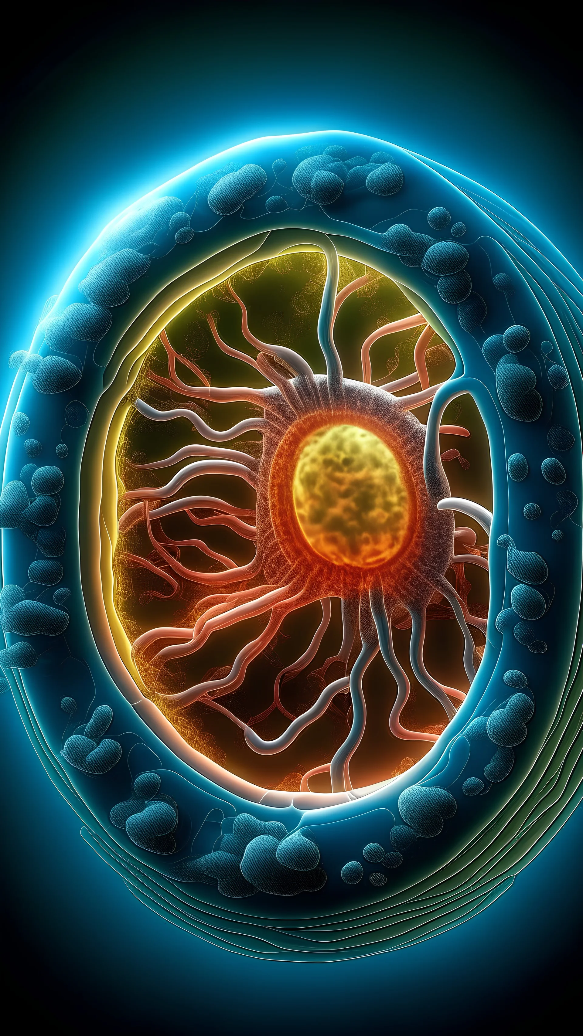
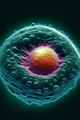
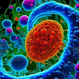
imagen de una celula procariota por dentro
3 years ago
Model
Kandinsky 2.2
Guidance Scale
7
Dimensions
2880 × 5120
Similar
@yesica
Prompt
3 years ago
Model
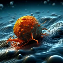
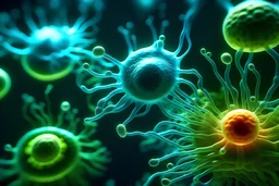
Kandinsky 2.2
Guidance Scale
7
Dimensions
2880 × 5120
Similar
© 2026 Stablecog, Inc.