@Shobu
Prompt
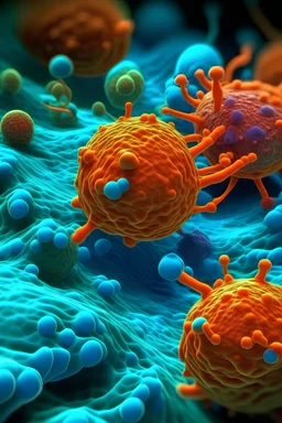
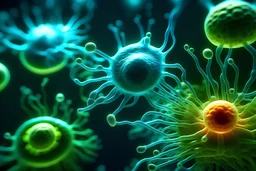
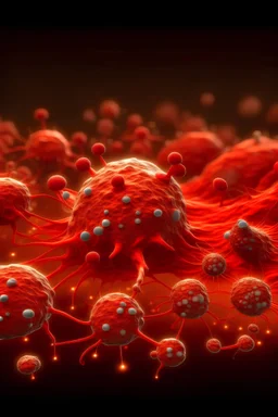
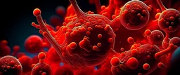
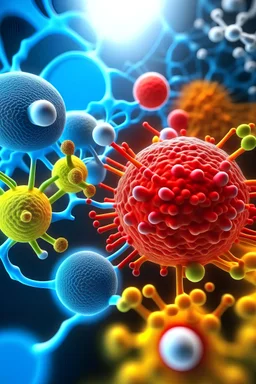
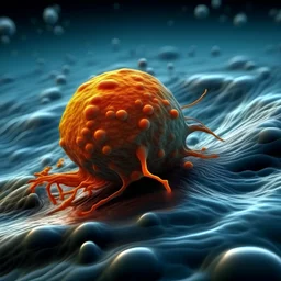
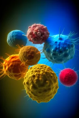
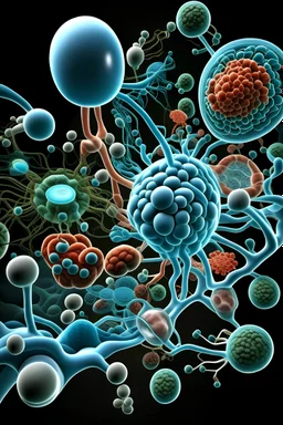
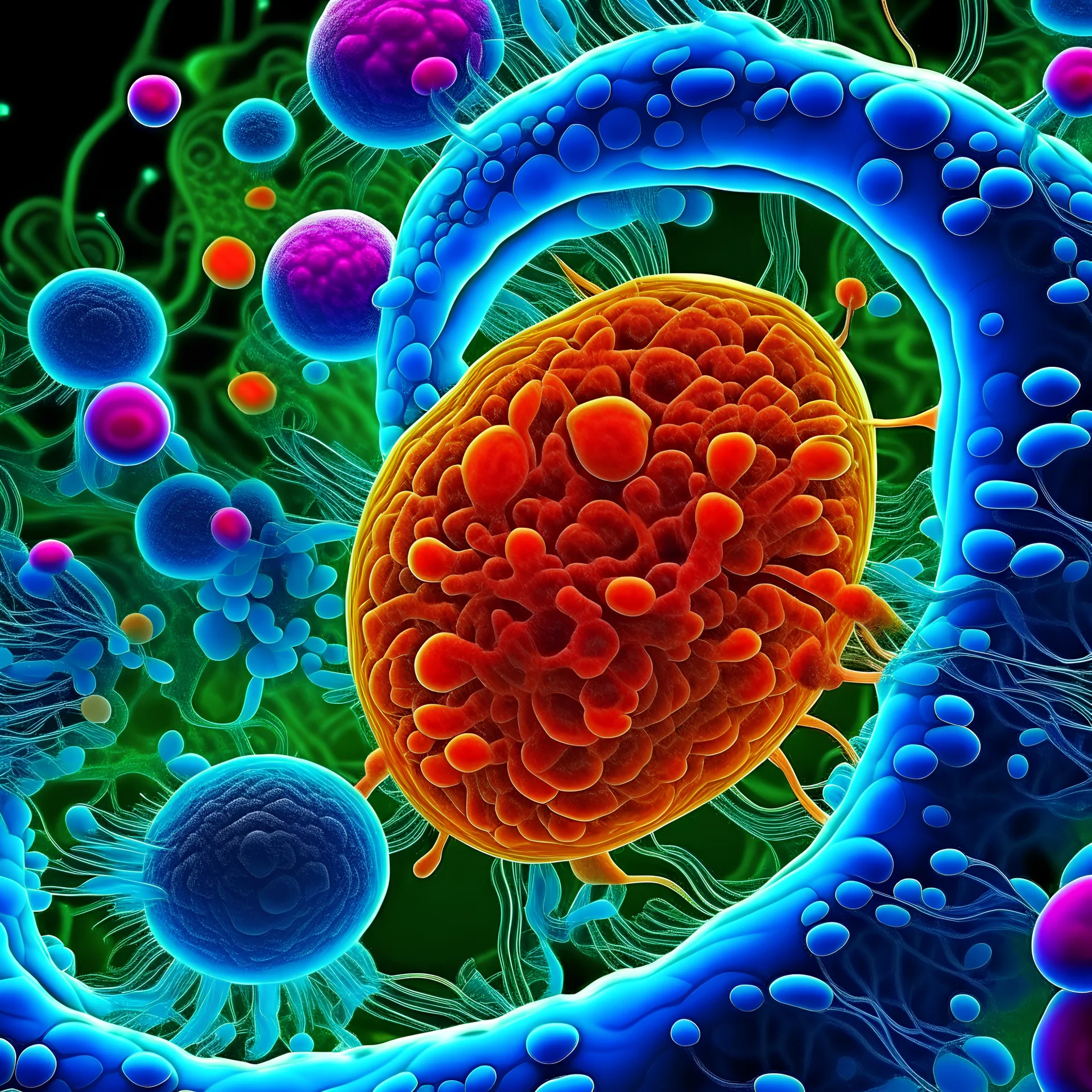
cellules eucaryotes adhérentes vues au microscope pendant la division cellulaire, pas d'animation, photo réaliste, cellule cancéreuse au milieu de cellules saines. Photo nette et très détaillée en 4K, grand angle, nette, vibrante, éclatante.
3 years ago
Model
Kandinsky 2.2
Guidance Scale
7
Dimensions
4096 × 4096
Similar